I'm an international teacher in Abu Dhabi. I am seeking new ways to support teachers. I am the face behind the Laney Lee science resources that you love!
Let me be real: teaching microscopy labs in middle school can feel intimidating. Between managing microscopes, stains, glass slides, and students who want to touch everything, it can quickly turn into chaos—especially if you don’t have a dedicated lab space.
That’s exactly why I started creating science labs that are simple, engaging, and realistic for everyday classrooms.
This Onion and Cheek Cell Stains Lab is one of my favorite introductory microscope labs because students get to observe real plant and animal cells with their own eyes. Instead of just memorizing diagrams from a textbook, they prepare wet-mount slides, stain specimens, and compare the visible structures of onion cells and cheek cells under a microscope.
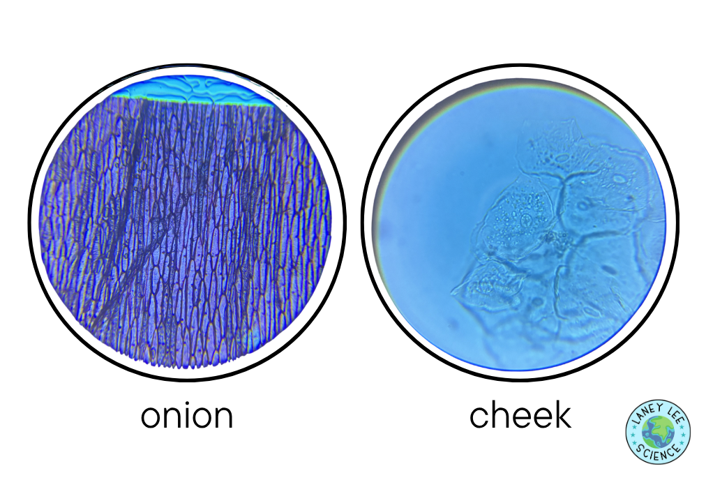
Not only does this activity align beautifully with NGSS MS-LS1-1 and MS-LS1-2, it also helps students develop critical lab skills like slide preparation, microscope use, scientific sketching, and evidence-based comparison. Students quickly notice that onion cells appear organized and rectangular because of their cell walls, while cheek cells are more irregular and flexible. Suddenly, “plant vs. animal cells” becomes something tangible and memorable instead of abstract vocabulary.
And the best part? This lab is surprisingly manageable with simple materials and basic classroom microscopes.
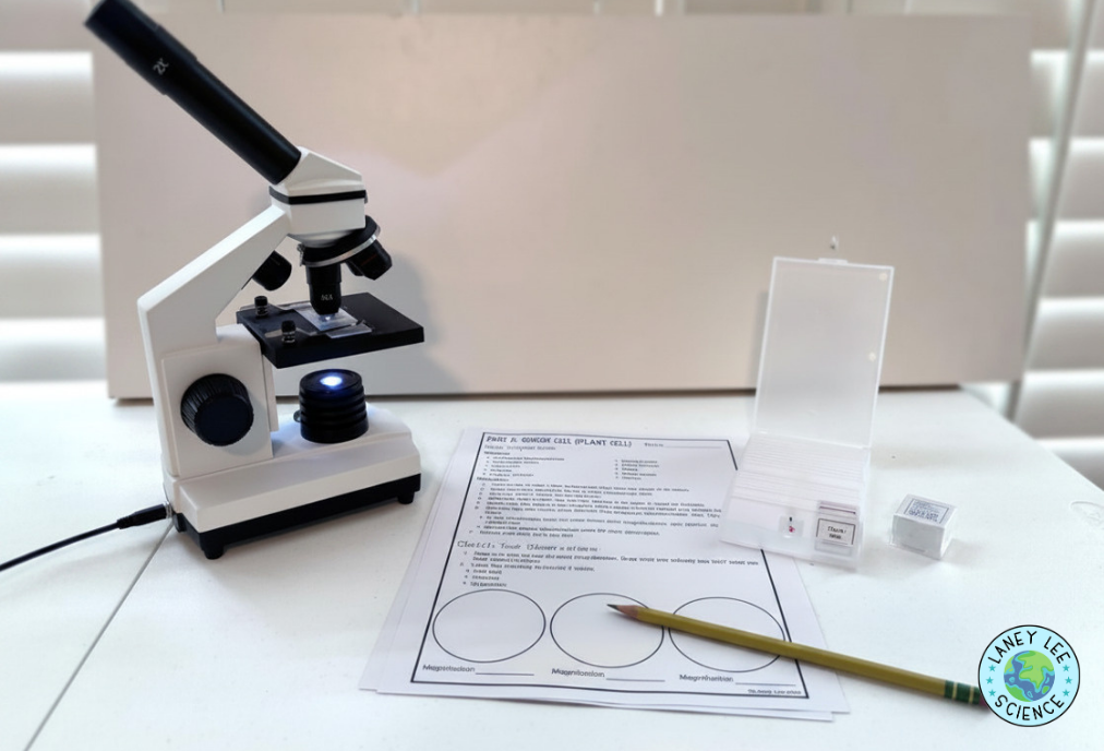
Students peel a thin layer of onion epidermis and place it onto a microscope slide. After adding iodine stain and carefully placing a coverslip, they observe the cells under multiple magnifications and sketch what they see.
Part B: Cheek Cells (Animal Cells)
Students gently collect cheek cells using a toothpick, smear the sample onto a slide, and stain it with methylene blue. They then observe the cells under different magnifications and compare the structures to the onion cells.
Data Analysis & Comparison
Students compare the shape, organization, visibility of nuclei, and presence or absence of a cell wall in both cell types. They also analyze how magnification changes the level of detail and field of view.
LANEY'S TIPS FOR SUCCESS
Simplify if needed. You can get a lot of preprepared slides online if prep is too daunting!
Model slide prep slowly. Students are usually most nervous about coverslips and staining.
Use very small stain drops. Too much iodine or methylene blue can flood the slide and reduce visibility.
Have students practice focusing at low magnification first before moving higher.
Stress “draw what you actually see.” Students love to draw textbook-perfect cells instead of real microscope observations.
Prepare extra onion samples ahead of time if your class periods are short.
conclusion
The Onion and Cheek Cell Stains Lab gives students an authentic introduction to microscopy while helping them truly understand the differences between plant and animal cells. By preparing and staining real specimens, learners observe how cell structure relates to function and build foundational lab skills that will support future biology investigations. It’s visual, engaging, highly memorable, and one of the best ways to bring cell theory to life in a middle school classroom.